KratosEditor V5 Rhinoplasty Architecture
4 Core Rhinoplasty Technologies Built for Surgeons
Patented clinical toolset that elevates patient consultations while delivering exact millimeter and degree targets to the operating room.
TipProj • DorsalHeight • TipDev • Angles
Multi-Dimensional Rhinoplasty Biometrics: Projection, Dorsal Height, Deviation & Angles
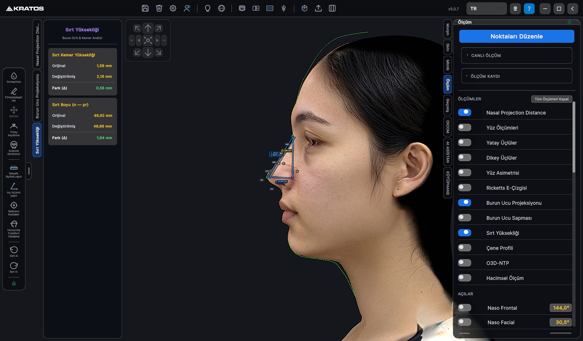
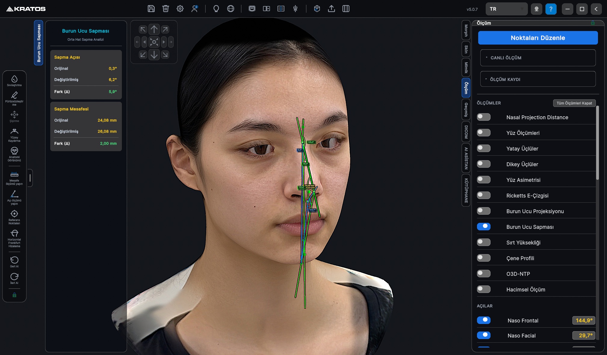
Real-time Pre-Op (Original), Post-Op (Modified), and Delta (Δ) clinical measurement suite: Nasal Dorsum Length (n–pr), Alar-to-Tip Projection (al–pr), and Tip Projection Ratio; perpendicular Dorsal Hump Height (mid_nasal to n–pr chord in mm); frontal-plane Tip Deviation Angle (°) and perpendicular midline offset (mm) relative to Nasion–Menton; plus Nasofrontal, Nasolabial angles and Ricketts E-Line balance.
Tip Projeksiyon Oranı (al–pr / n–pr)
Dorsal Yükseklik & Uzunluk (mm)
Frontal Tip Deviasyon (° & mm)
Nasofrontal & Nasolabial (°)
Ricketts E-Line
AnalysisVisualizer.NasalProj
6-Level Sagittal Nasal Profile & Dorsal Resection Analyzer
Superimposes Pre-Op (Green) and Planned Post-Op (Orange) sagittal profile curves in real time. Quantifies exact anterior-posterior dorsal reduction or radix/tip augmentation in millimeters across 6 standardized levels along the nasal bridge, complete with an interactive Orbitale (Or) A–B–C reference triangle.
1. Radix (10%)
2. Rhinion (27%)
3. Midvault (44%)
4. Supratip (61%)
5. Pronasale (78%)
6. Infratip (95%)
Nasal_v1.4 & Symmetry Macros
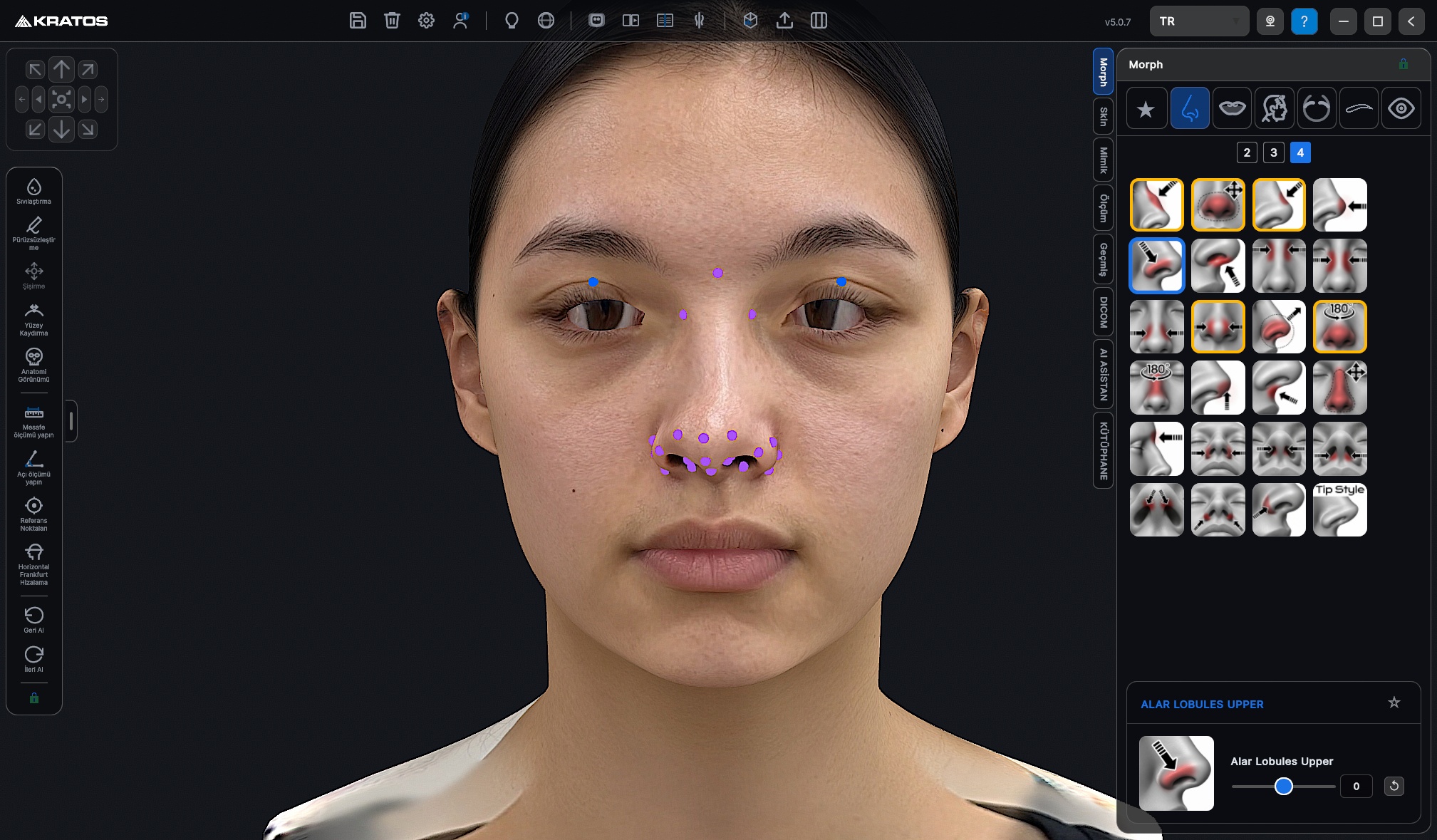
24 Specialized Rhinoplasty Controls, Auto-Basal View & Symmetry Macros
Granular control over Bony Pyramid Lateral Osteotomy (SideWallUp), Midvault Spreader zone (SideWall), Bulbous/Boxy Tip Slimmer, Supratip Break, Alar Base Weir Resection (with automatic 45° basal camera switching), Columella Show, and Nostril Length. Includes 1-click NoseBridgeSym and NoseTipSym macros for deviated revision cases.
NoseBridgeSym
NoseTipSym
AlarBase Weir
SideWall Osteotomy
Clinical Nose Library
X-Ray Lens • DICOM • 1:1 STL Splint
X-Ray Subcutaneous Lens, CBCT Probe & 1:1 3D Printable Nasal Splint
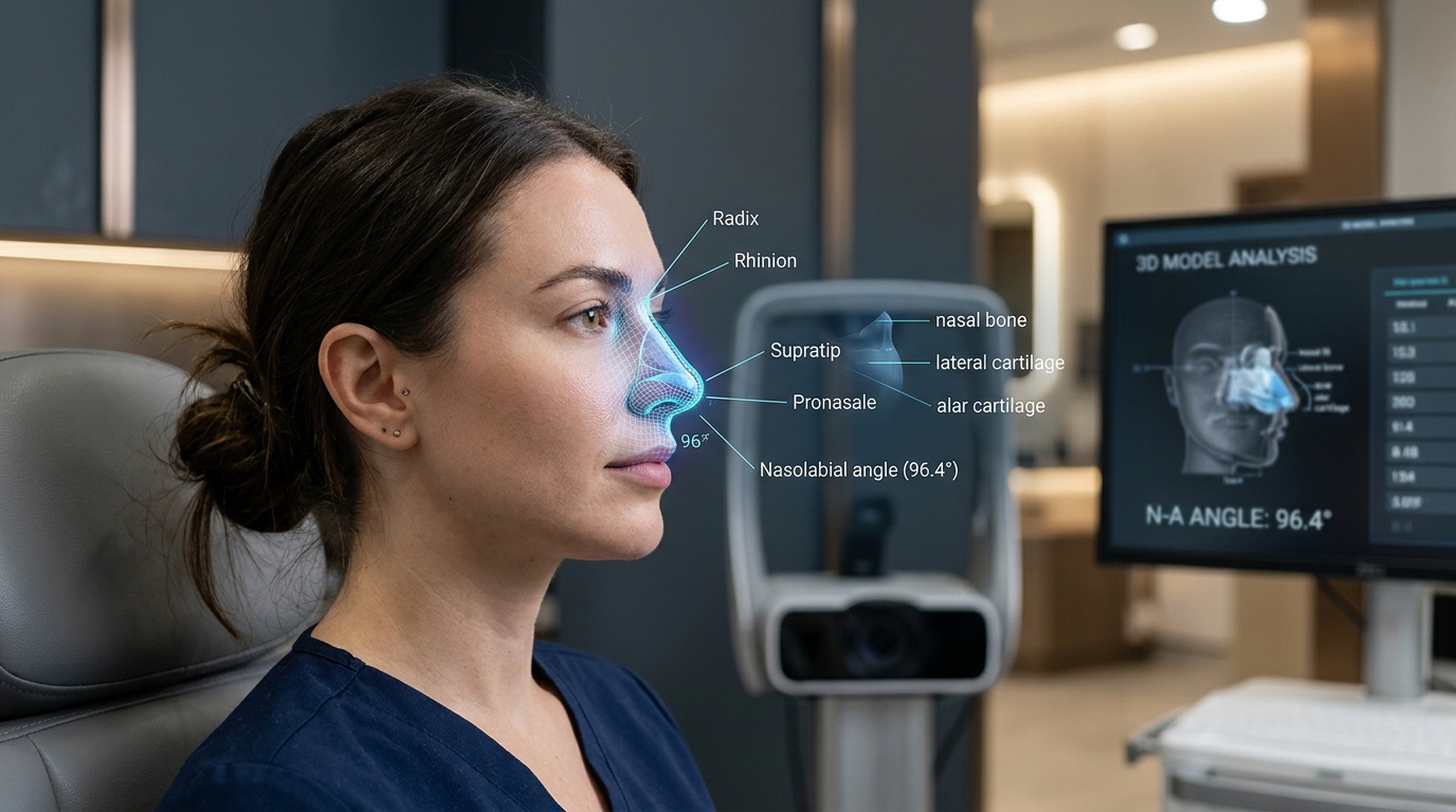
Inspect underlying upper/lower lateral cartilages and nasal bones through a movable 3D X-Ray lens. Register CBCT/DICOM scans with the 3D optical surface to probe exact skin-to-bone tissue thickness in millimeters, and export a 1:1 real-world millimeter hollow nasal splint (2.5 mm wall thickness) in STL/OBJ/PLY for in-clinic 3D printing.
X-Ray Cartilage Lens
CBCT Skin Depth (mm)
1:1 Hollow Splint STL
Nasofrontal & Nasolabial °